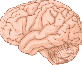
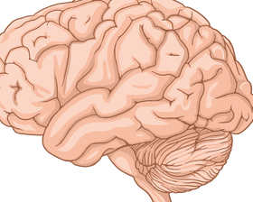
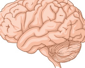
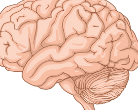
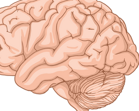
Modern neuroscience uses complementary tools to study brain structure, electrical activity, metabolism and function.
How It Works
MRI provides detailed structural images; CT uses X-rays and is especially useful in urgent assessment; PET can measure selected metabolic or molecular activity.
Key Science
EEG records electrical activity from the scalp, while fMRI measures blood-oxygen-related changes associated with neural activity.
Why It Matters
No single technique shows everything about the brain. Clinical interpretation depends on symptoms, medical history and the question being investigated.
Quick Answer
This topic is part of modern neuroscience and is best understood through interacting biological systems, individual variation and the limits of current evidence.
FAQs
Is one brain region responsible for everything?
No. Complex brain functions generally depend on distributed networks that communicate across regions.
Can brain function change over time?
Yes. Development, learning, experience, injury and aging can influence neural structure or function.
Sources
NCBI Bookshelf · NIH NICHD Neuroscience · NIH BRAIN Initiative
Converse Video
Official Converse
For current models, materials, availability and sizing, check Converse.com.
Converse